animal cell under microscope diagram
Since objects viewed under the microscope are. Animal Cell Under Microscope.
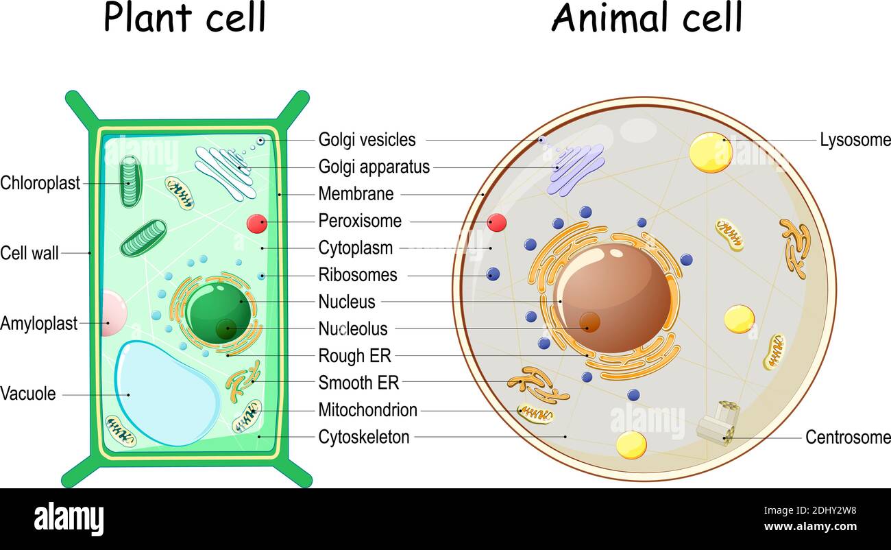
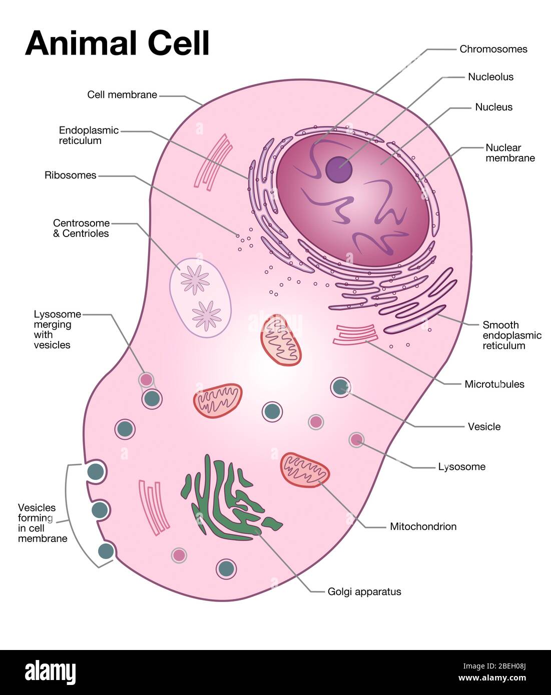
Animal Cell Structure Hi Res Stock Photography And Images Alamy
A cell is the smallest functional and structural entity of life that it is easier observing animal cell under light microscope.
. A sample picture of each a plant animal. Pin on R Cell Assignment. Diagram Of An Animal Cell Under A Microscope - animal cell microscope slide wallpapersskin - Answer the following questions in your exercise book.
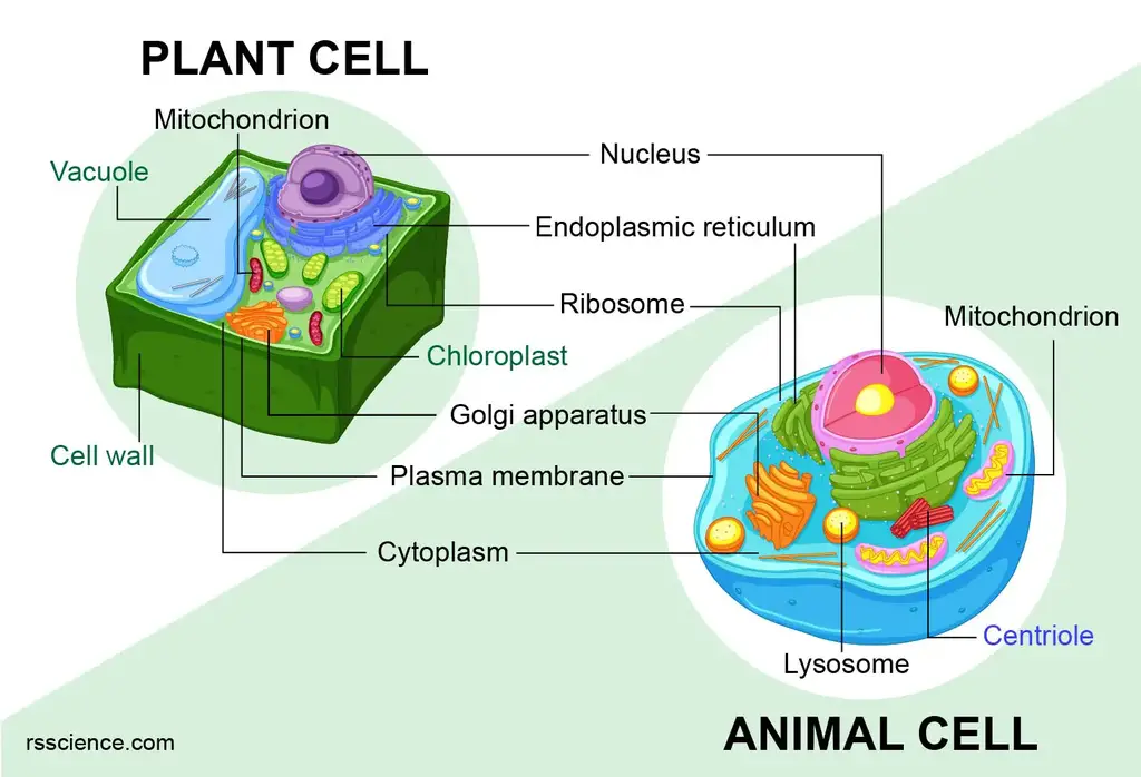
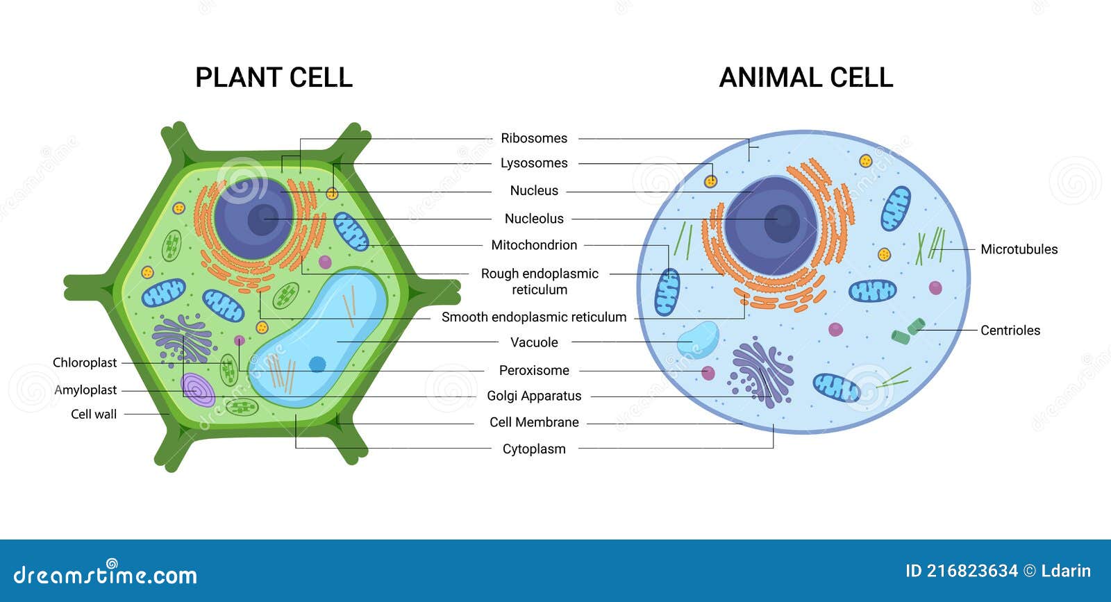
The organelles and structures which are common to both plant and animal cells are- Plasma membrane. Just under the rigid cell wall is the more fluid cell membrane. Students will discover that their skin is made up of cells.
So hair is an epidermal down growth embedded into the dermis or hypodermis of the animals skin. A typical animal cell is 1020 μm in diameter which is about one-fifth the size of the smallest particle visible to the naked eye. Diagram of parts of a microscope.
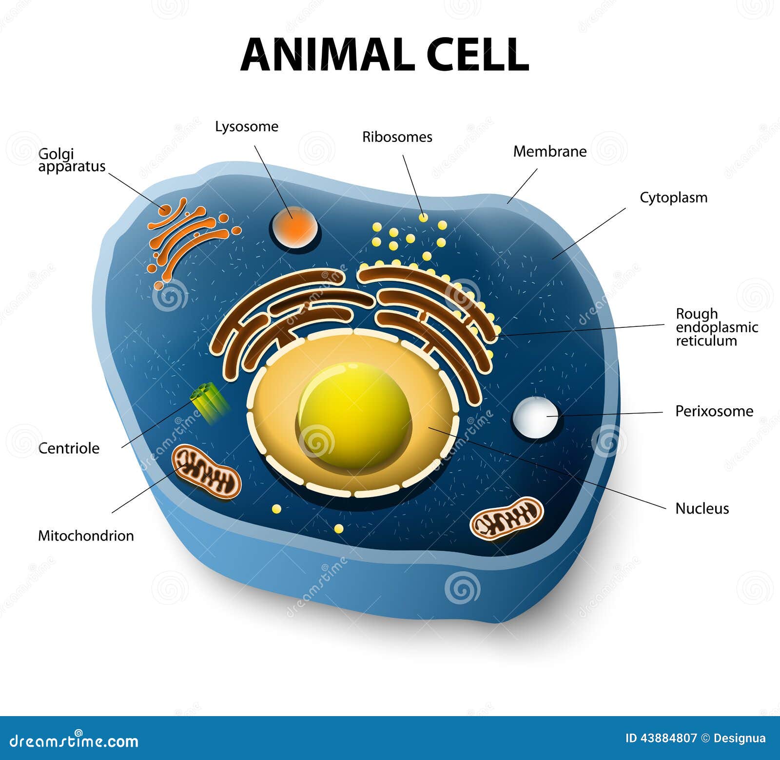
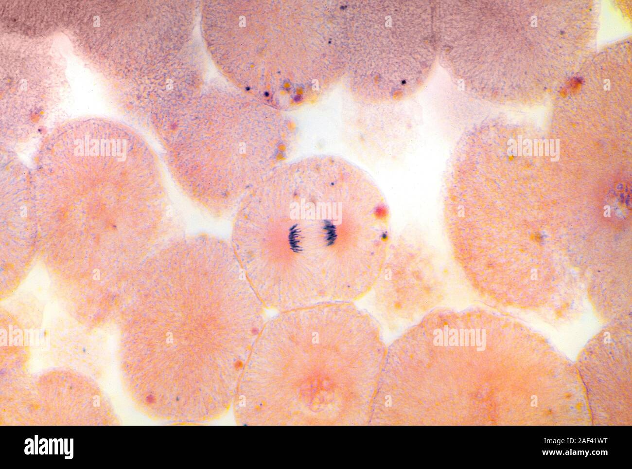
Animal Cell Diagram Under Light Microscope. Animal cell as seen under electron microscope. Most of the cells are microscopic hence they can only be seen under a microscope in order to study their anatomy.
Most of the cells are microscopic in size and can only be seen under the. More images for diagram of animal cell under light. Print the scripts for animal and plant cells.
May 15 2019 animal cell structure diagram model animal cell parts and. But at the same time it. But at the same time it.
This shows a generalized animal cell under a light. Hair under microscope. To make observations and draw scale.
Animal Cell Diagram Under Light Microscope. Animal cell under the microscope. Students will discover that onions are made up of cells.
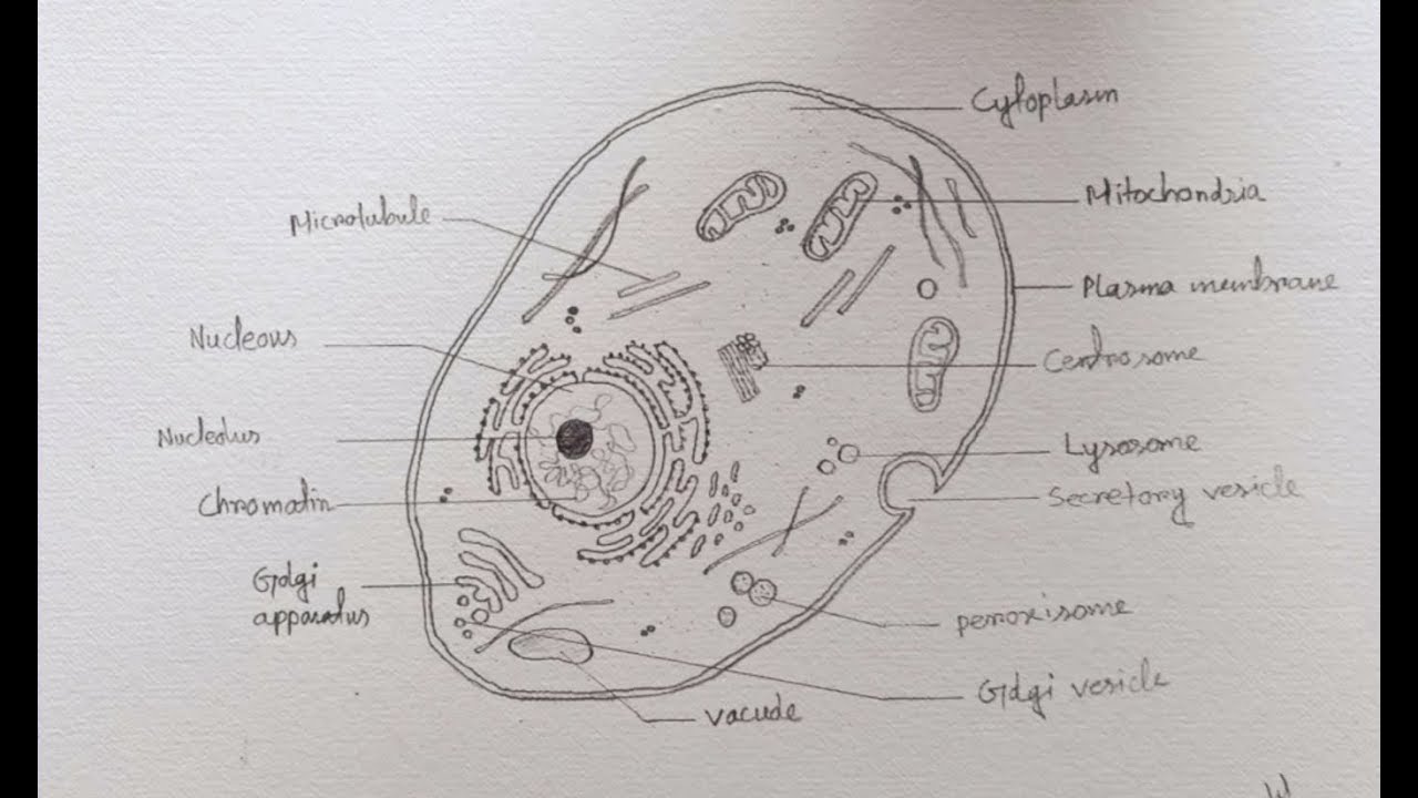
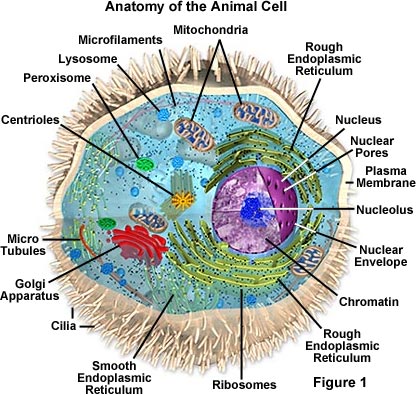
Nucleus Mitochondria Endoplasmic reticulum Golgi apparatus The chief. You will find two main parts in hair a cylindrical. Though this animal cell diagram is not representative of any one particular type of cell it provides insight into the primary organelles and the intricate internal structure of most.
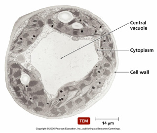
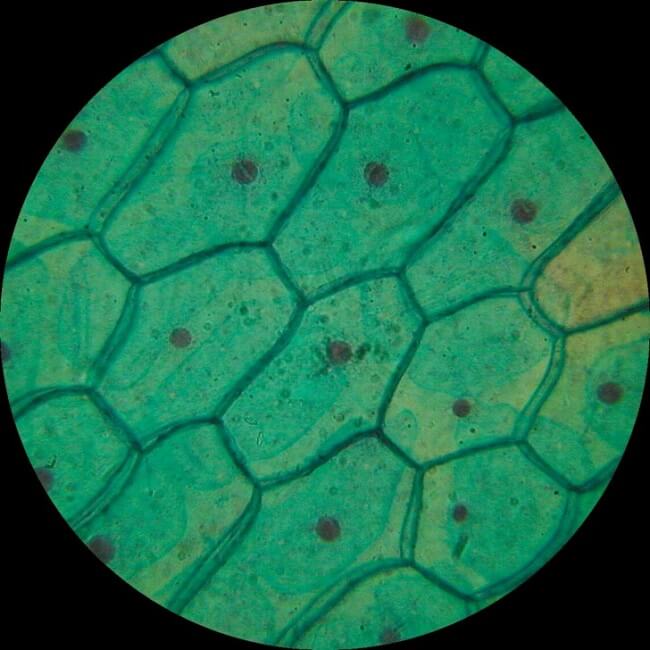
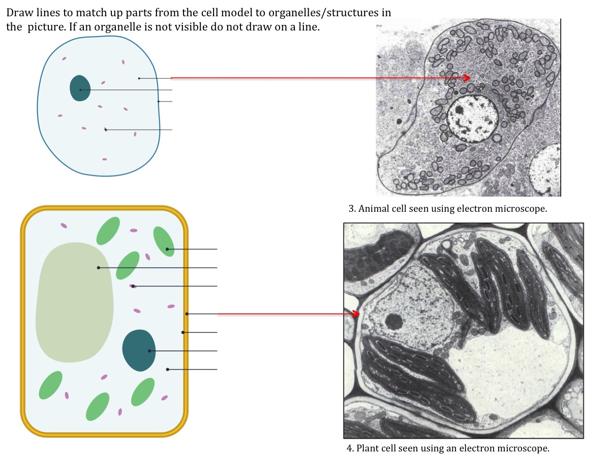
14 can you distinguish features of the cells in fig. Students will observe onion cells under a microscope. When looking under a microscope the cell wall is an easy feature to distinguish plant cells.
Diagram Of Animal Cell Under Microscope.

Q14 Draw A Large Diagram Of An Animal Cell As Seen Through An Electron Microscope Label The Parts Brainly In

Gce Cie Biology Animal And Plant Cell Structures And
Plants Vs Animals Bronco Biology Exploring The Cell
What Are The Parts Of An Animal Cell And Its Functions Quora
Animal Cell Microscope Stock Illustrations 1 941 Animal Cell Microscope Stock Illustrations Vectors Clipart Dreamstime
Animal Cell Diagram Hi Res Stock Photography And Images Alamy

Animal Cells Under Microscope Stock Photo Picture And Royalty Free Image Image 57823181

Plant Cell Diagram By Russell Kightley Media
Animal Cell Microscope Hi Res Stock Photography And Images Alamy
Amazing 27 Things Under The Microscope With Diagrams

Eukaryotic Cells Under The Microscope 2 1 6 Ocr A Level Biology Revision Notes 2017 Save My Exams
Animal Plant Cells Stock Illustrations 374 Animal Plant Cells Stock Illustrations Vectors Clipart Dreamstime
How To Draw Diagram Of Animal Cell Easily Step By Step Youtube
Cell Structure Teaching Resources The Science Teacher
Pinkmonkey Com Biology Study Guide Chapter 3 Cell The Basic Unit Of Life

Animal Cell Diagram By Russell Kightley

Animal Cell Cell Diagram Plant Cell Diagram Animal Cell
Animal Cell Structure Function Diagram And Types
Molecular Expressions Cell Biology Animal Cell Structure